Book Free Session on WhatsApp or Call   +91  77000  04970

Record taking for aligner cases

Case history, X rays (OPG & lateral cephalogram), intraoral and extra oral photographs, impression of intraoral structure.

Case history –

Patient’s complete details on case paper

Medical history

Chief complain - The patient chief complaint should be recorded in his or her own words. It helps the clinician in identifying the desires and priorities of the patient.

Intra oral examination-

·       Mention all teeth present

·       Caries present

·       Grossly carious tooth

·       Tender on percussion

·       Missing teeth

·       Gingiva

·       Stains and calculus present

·       Overjet

·       Overbite

·       Molar relation on both sides

·       Crossbite

·       Midline shift

·       Tongue size

Extra oral examination- 

·       Swelling

·       Facial asymmetry

·       Facial profile

·       Competent lips

·       Nose

·       Facial asymmetry

OPG – Orthopantomogram is one of the most popular records in orthodontics diagnostic phase, it provides important bilateral dental and skeletal information.

Lateral cephalogram –Lateral cephalogram is used primarily in orthodontic diagnosis and treatment planning, particularly when considering orthognathic surgery.it is useful record to prior to treatment and can be used during treatment to assess progress. It is used to assess the etiology of malocclusion to determine whether the malocclusion is due to skeletal relationship, dental relationship or both.

Intraoral & extra oral photographs-

Intraoral photographs should take-

·       At occlusion, from front

·       Right side occlusion

·       Left side occlusion

·       Upper arch

·       Lower arch

Extra oral –

·       Full face at rest

·       Full face smiling

·       Turn patient completely 90 degree, at rest and smiling.

·       Turn patient 45 degree, at rest and smiling (make sure half of eyebrow of other side should be visible.)

·       While taking extra oral picture, only head neck portion should be visible.

·        Both the ears should be clearly visible.

·       Space should not be left in the picture above the patient’s head.

Impression of intraoral structure

 In Impression of maxillary and mandibular arches it should cover the all teeth present in jaws and structures like labial frenum, labial vestibule, buccal frenum buccal vestibule, hamular notch and in mandible labial frenum, labial vestibule, lingual frenum, buccal frenum, buccal vestibule & retro molar pad. 

Absolutely not.  Consultation can be done by only dental mirror and probe.

For intraoral & extraoral photographs a good quality back camera of cell phone are also useful & in case if you have the DSLR camera then it is more useful for orthodontic photographs, but it is not compulsory.

For impression of maxillary & mandibular arch impression trays, rubber bowl & mixing spatula is enough.

When taking impressions for aligner treatment planning and record taking, the goal is to accurately record the intraoral structures. The interference dental assistants experience while taking impressions is patient discomfort. This can present itself in several form, including fear, distress and gagging. By avoiding patient discomfort, dental assistants can create predictable and accurate result by using VPS (vinylpolysiloxane) impression material, allows for maximum patient comfort and the most efficient impression taking procedure.

Firstly using the putty, knead and fold the catalyst and base. Load the putty around the tray making sure you have an equal amount of material.

Make concavity where the teeth should be with your finger before tray placement.

Add the correct flow wash right on top of putty and seat into patient’s mouth.

Seat the tray into the mouth. When the time is complete, remove the impression.

Yes, you can take the alginate impression for initial diagnostic record in aligner cases. In orthodontics impression gives a negative imprint of the tissues and by filling it with dental stone a positive cast is made that can be removed after model material has set.

Manipulation of alginate – the recommended water powder ratio should be used. In general it is 38ml of water per 16gms of powder but slight variation can be presented depending on the manufacture. The weighed power is include into the water by carefully mixing with the spatula. Care should be taken that air is not incorporated into the mix by spatulating the mix against the side of the bowl while using the vigorous figure of 8 motion till a smooth creamy mix is obtained.

Maxillary impression –

Operator position at 11 o’clock position

Patient seated such in a way that the occlusal plane should parallel to the floor

A bead of material may be placed in the palatal surface of the patient just behind the incisors, to remove the trapped air and to ensure the faithful reproduction of palatal tissue.

Use the thumb and index finger of the free hand to retract the check.

Once inserted straight the impression tray so that the tray is in line with the patient’s mid face.

Start to apply light pressure upwards till the light resistance is felt.  

Mandibular impression-

Operator position at 7 o’clock

Patient seated in a way that the occlusal plane should parallel to the floor.

Use the thumb and index finger of the free hand to retract check.

Grasp the handle of the tray and rotate the tray into mouth by using the front of the tray to deflect the other check.

Once inserted straight the impression tray so that the tray is in line with the patient’s midface.

Note the position of the tray in relation to the anterior.

Using the index finger of both hands press the tray downwards

Manipulate the patient’s cheek and lip to create a more detailed anatomy of the facial and vestibular area.

Ask the patient to elevate the tongue and then move it from side to side.

Allow the material to set and ten remove. Using a firm grip on the tray handle, use a sideways lifting motion to remove the tray.

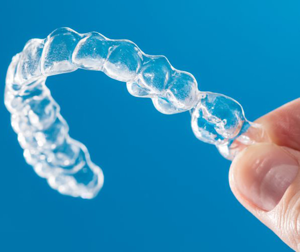
Intra-oral photographs: The major purpose of intraoral photographs is to enable orthodontist:

          To view the hard and soft tissue at clinical examinations.

          To report hard and soft tissue condition as they exist before treatment.

Requirements for intra oral photographs are –

Quality, standardized intra oral prints in colour

Patient dentition oriented perfectly in all three planes of space

One frontal view in maximum intercuspation.

Two lateral view right and left.

Optional two occlusal view maxillary and mandibular.

Free of distraction – cheeck retractors, labels, and fingers

Quality lighting which shows anatomical contours and makes image free of shadows.

Tongue retracted

Free of saliva and bubbles

Clean dentition & photographic records were obtained by setting up a neutral and plain background.

Guidelines - 

It is important to get permission from the patient before taking photographs.

Patient should be seated, bent back slightly in the chair.

Height of the chair should be adjusted such that the patient’s head is lower to that of photographers.

A stable position of photographer is mandatory (since camera is handled and not placed on tripod)

The upper arm held against upper part of body with the left hand supporting front of lens.

The eye is not pressed against the eyecup but slightly in back of it. The other eye to be open.

Photographer should be supported by outside edge of the patient chair in order to find secure comfortable position.

Select magnification ratio according to the desired frame and focus by moving camera back and forth.

20 cm is good distance to start testing a camera’s ability to take sharp anterior intra oral photographs on the manual setting.

Different view of photographs-

Frontal view – the frontal view is must as it details the appearance of teeth as seen by patient, parent and general public. This view is prefer in particular for general purpose and orthodontics, this is taken in landscape orientation with the teeth in occlusion filling the frame with the occlusal frame horizontal and bisecting the picture. Large ends of larger retractor should be used. Assistant should hold both retractors, pulling all the soft tissue laterally and forward, this makes it easier for the patient to bite together in occlusion and pulls the soft tissue away from teeth. The midlines if they should be at the centre of frame. Mouth, adequate depth of field is required, so it is must to focus on lateral incisor or mesial of canine to ensure that maximum no. of teeth are in focus. The centre of image is the contact point of upper central incisors.

Buccal view - patient told to close in maximum intercuspation. Occlusal plane should be horizontal. Mirror is always required. A proper wide lateral mirror is inserted distal to last tooth turned outside as far as lips and check will stretch should not on gingiva. Patient can be asked to hold the mirror. Angle of camera should be adjusted so that lens is perpendicular to the buccal surface of posterior teeth. The centre of photograph and focus point are around second premolar or first molar, depending upon framing of image.

Occlusal view maxillary – images should extend should extend from just in front of incisors to at least distal surface of first molars and ideally to include all erupted teeth. There should be no direct view of incisor. Patient position- head tilted backward so that photographer does not have to twist excessively, instruct patient fully open mouth. Palatal mirror rest on distal aspect of last molar and turned down until it touches the incisors. Centre of photographs is a cross section of sagittal plane with the connection line between second premolar running horizontally in the middle of image.

Occlusal view mandibular – palatal mirror inserted with the border end so that mirror rests on distal aspect of last molars, it is turned upward with mouth wide open until it touches the incisal edges of upper incisors. The patient is advised to raise the tongue & breath through nose .centre of image should be at the intersection of the sagittal plane with the line crossing second premolars positioned horizontal in the centre of image.

Extraoral photographs-

Requirements are –

Quality standardized facial photographs either in black and white in colour

Patient head should place accurately in all three planes of space and in Frankfort horizontal plane

One lateral view facing to the right serious expression lips closed tightly to reveal muscle imbalance and disharmony

One anterior view serious expression

Optional one lateral view and or one anterior view with lips apart

Optional one anterior view smiling

Background free of distraction

No shadows in background

Ear exposed for purpose of orientation

Eyes open and looking straight ahead, glasses removed.

Positioning of patient- both patient & clinician need to be positioned correctly in standardized manner. If there is height difference between patient and clinician any one of them can stand on a platform to raise them to appropriate level camera level at the middle of face. In extraoral photography attempt should be made to focus on the patient’s lower eyelid to ensure from tip of nose to ear of the patient falls within the depth of field

Frontal view - portrait view with the frame extending to just above the top of head and lower frame line around the larynx. Photographs should be symmetrical with the interpapillary line parallel to floor. A focusing screen with the grid is very useful. Patients assumes a natural head position and looks straight ahead into the camera. Camera positon middle of face and in portrait format. Space should be left on all sides of photographs. 

Frontal at rest- teeth in maximal intercuspation with the lip closed even if this strains the patient in case of lip incompetence. This photographs shows as clear documentation of lip strain and its aesthetic affect.

Frontal dynamic smile- smiling picture demonstrate the amount of incisor smile (percentage of maxillary incisor display on smile) as well as excessive gingival display.

Profile view- usually only one profile (right profile matching up with the lateral cephalogram) is taken. For a patient with facial asymmetries both right and left profile should take. Frame extending to above the top of head in front of nose and below chin. Back of head is not necessarily required, the remaining free space should be in front of profile. Patient assumes a natural head position & look straight ahead in relaxed manner and lips also relaxed. Frankfort plane is horizontal and parallel to the horizontal frame of photographs.

Three quarter profile- useful in examination of midface deformities in surgery of jaw. Portraits should be taken in such way that the sagittal plane of patient and optical axis of camera are approx. 45degree to each other.

Dental cheeck and lip retractors

“C shape” retractors- The “c shape” of the retractor moves the soft tissues of the lips and cheeks up and down, so that all kinds of dental work can be presented in more detail and transparency.

“V shape” retractors- The “v” shape of the retractors will be able to stretch more the lip and cheeks outwards, where we can see the side of the teeth better.

Generally, in orthodontics it is better to use the “v shape” because by pulling the soft tissues of lips and cheeck outwards, the lateral teeth are more exposed as carriers of the occlusion.

Many types of mirrors have been used ranging from front-silvered mirrors to highly polished stainless steel mirrors.

Front silvered mirrors gives the best quality image and light distribution, no ghost image or double layering.

Light reflection on front silvered mirrors brightens, bright image as an end result.

Long-handle mirrors- it is preferred to use “long-handle” mirrors as they allow better control and handling by the clinician during the occlusal shots. You can find various sizes for use with different patients depending on age and mouth-opening size, but generally, the “medium” sized mirrors would be fit for use with most patients.

Yes wax bite is needed for Aligner cases, as it gives the exact occlusal relationship for proper guidance, it gives accurate cusp to fossa relation of maxilla and mandibular teeth or accurate angle’s malocclusion.

Method of bite registration

The centric position is check and the desired degree of activation decided.

The patient is then educate to bite in the desired position by giving him a mirror.

The wax is softened in water bath and adapted.

The patient is instructed to bite into the accurate position.

After the wax has hardened properly, it is removed and chilled.

The models and wax bite are articulate & kept aside for future reference or courier send for aligner fabrication.

Intraoral & extraoral examination-

Intraoral examination-

There are two type -

·       Soft tissue examination

·       Hard tissue examination

Soft tissue examination-

Gingiva- Anterior marginal gingivitis usually occurs in mouth breathing and open mouth habit. While generalized marginal gingivitis is associated with poor oral hygiene.

Frenum – A persistent and thickened labial frenum may be present in 25% of children at age of 8 years.

Palate – should be examined by palpation and visually. Asymmetrical contour are usually caused by unerupted teeth.

Tongue – is a combination of muscles around which dentition is shaped and melded.

Its size: large tongue cause generalized diastema.

Its action: forward positioned tongue causes open bite, backward positioned tongue may cause anterior crowding.

Hard tissue examination                     

Number of teeth

Teeth present & erupted

Teeth present & unerupted

Extracted teeth

Congenitally missing teeth

Extra teeth

Condition of teeth

          Carious or sound

          Non-vital or vital

          Teeth with extensive restoration

          Broken teeth

          Malformed or fused teeth

Proportion of tooth size to arch size

          Crowding occurs where teeth are large compared to jaw size.

Position of teeth

          Any of individual teeth malposition.

Malposition of individual teeth 

          Labioversion

          Supraversion

          Mesioversion

          Torsoversion

          Linguoversion

          Infraversion

          Distoversion

          Transversion

Malrelation of the dental arches

Anteroposterior malrelationship

·       Class I

·       Class II

·       Division 1

·       Division 2

·       Class III

Vertical malrelationship

          Deep overbite

          Edge to edge bite

          Open bite

          Transverse malrelationship

Posterior cross bite (unilateral bilateral or telescoped bite)

Midline shift

Extraoral examination

A.Shape of head-

·       Mesocephalic

·       Dolicocephalic

·       Brachycephalic

B.Facial form-

·       Mesoprosopic

·       Euryprosopic

·       Leptoprosopic

C.Facial profile

·       Straight profile

·       Convex profile

·       Concave profile

D.Facial Divergence –

·       Anterior divergent

·       Posterior divergent

·       Straight or orthognathic

E. Examination of lips-

·       Competent lips

·       Incompetent lips

·       Potentially incompetent lips

·       Everted lips

F .Examination of chin –

·       Mento-labial sulcus

·       Mentalis activity

·       Chin position and prominence

Either it get by online /soft copy format – consent form will be available on our website we can share you a link via mail, where you can  download the consent form with signature of patient & doctor and scan it & mail it or submit online.

Hard copy format – will be sending consent form via courier you can take sign of patient & doctor & courier it back to us.